Welcome to International Thyroid Eye Disease Society !

Surgical Therapy

Timing of surgery

Surgical rehabilitation for TED is considered during the inactive phase following several months of stable measurements and controlled thyroid status (see Fig). Surgery may be necessary during the active phase when vision is threatened and includes orbital decompression for refractory DON or tarsorrhaphy and lid-narrowing procedures for corneal exposure.

Surgery for TED is staged sequentially:

1.Orbital decompression

2.Strabismus repair

3.Eyelid surgery

Rundle curve and surgical timing

Orbital Decompression

This is used for dysthyroid optic neuropathy, proptosis, and chronic orbital congestion. Decompression involves bone wall and/or fat removal, depending on the indication, the targeted tissue, and the desired amount of proptosis reduction.


Optic nerve apical compression (DON) by expanded EOM is relieved by out-fracture of the medial orbital wall into the posterior ethmoid sinus and the medial floor into the maxillary sinus (Fig:  Coronal CT showing LE apical crowding pre and post medial wall decompression). Even significant or longstanding vision loss may be reversed by an effective decompression. Adjunctive CS or RT may be offered to avoid postoperative deterioration in vision because of active disease with progressive muscle expansion.48


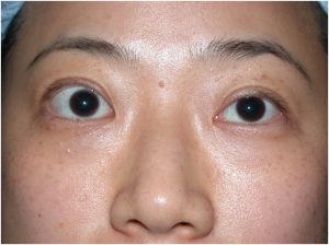
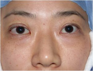
Proptosis reduction during the inactive phase is the most common indication, to improve comfort and appearance (Fig. Top shows RE proptosis and lid retraction; centre shows enlarged EOM and fat; bottom post decompression and lid narrowing). A swinging eyelid approach allows access to the lateral wall, floor, and medial wall, and any combination of bone wall removal and fat removal may be chosen, depending on the relative contribution of fat versus muscle tissue expansion. Specific complications are associated with each of the walls removed. Cheek numbness and globe inferior displacement may occur with floor decompression, oscillopsia may occur with lateral rectus/temporalis muscle apposition following lateral wall decompression,53 and sinusitis and ethmoidal nerve anesthesia may arise following medial wall decompression.


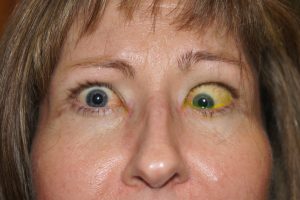
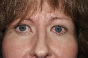
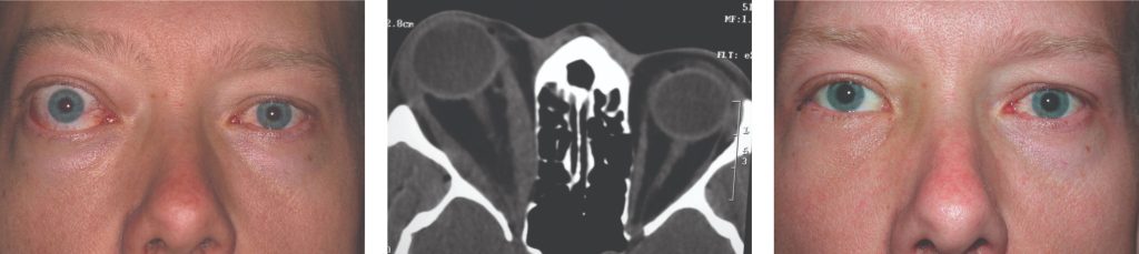
Reduction of long-standing orbital congestion is a rare indication. These individuals may have high CAS/VISA inflammatory scores, but the disease is inactive (false positive CAS) with stable VISA severity grades and poor response to GC. Improved venous drainage following surgery often results in a gratifying and immediate improvement in CAS/VISA I scores (Fig. shows inactive congestion on left and improvement after decompression on right, CLICK to enlarge).

RE Proptosis before and after decompression


Strabismus is a troublesome consequence of decompression surgery, particularly in cases with enlarged muscle and pre-existing strabismus (DON and congestive disease). It is rare in cases of fat-targeted disease.

Strabismus Surgery

Individuals with muscle-targeted progressive TED have a significant risk of developing muscle scarring, restriction, and strabismus with significant impact on QOL.


At the first symptoms of diplopia, combined radiotherapy and corticosteroid therapy might limit progression and preserve the greatest field of single binocular vision.


Strabismus and ductions can be assessed both in the office and by an orthoptist so that a temporary prism can be fitted and measurements repeated until stable for at least 6 months, when surgery may be offered. The orthoptist also measures fusional amplitudes and torsion to assist in surgical planning.


Most strabismus surgery for TED consists of recessions, often on adjustable sutures (Fig. shows active TED with strabismus and lid retraction LEFT, and post-operative alignment surgery and lid narrowing surgery RIGHT, CLICK to enlarge).


Inferior rectus recessions are prone to slippage and may induce lower lid retraction.  


Eyelid Surgery

Upper eyelid retraction is associated with enlargement and scarring of the levator complex and is treated with recession of the scarred complex (Fig. shows RE upper lid retraction LEFT, and post-operative posterior levator recession RIGHT, CLICK to enlarge). 

Lower eyelid retraction is mostly associated with proptosis, although secondary retraction from inferior rectus recession is well recognized. Orbital decompression with floor removal may accentuate upper lid retraction (as the upper lid remains tethered while the globe is relatively lowered), whereas lower lid retraction may be reduced.


In progressive disease, retraction is usually managed conservatively with topical lubricants unless the cornea is threatened, in which case a temporary tarsorrhaphy or levator release may be performed.

Management of TED Using VISA Classification